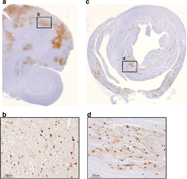
Figure 2.

Distribution of NP antigen in positive tissues of a quail intranasally challenged with H5N1/HP. a. Brain, 7 dpi. b. Positive staining in nucleus and cytoplasm of neurons and glial cells. c. Heart, 5 dpi. d. Positive staining in nucleus and cytoplasm of myocardiocytes.
