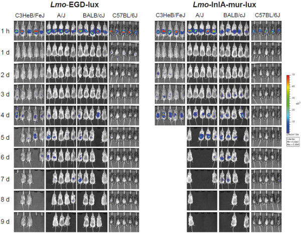
Figure 1.
Bioluminescence imaging (BLI) of listeriosis in different inbred mouse strains after oral infection challenge with Lmo-EGD-lux and Lmo-InlA-mur-lux. Ten female C3HeB/FeJ, A/J OlaHsd, BALB/cJ and C57BL/6J mice were intragastrically challenged with 5 × 10⁹ CFU Lmo-EGD-lux (left column) or Lmo-InlA-mur-lux (right column) and the progress of infection was assessed by BLI for 9 days. Bacterial luciferase activity was visualized in five mice per measurement using the IVIS 200 imaging system as described in Methods. Serial BLI data are shown for a set of five mice for a time period of 9 days p.i.. They are representative of two independent experiments each with a total of 10 mice per inbred mouse strain. Empty spaces indicate dead mice. The colour bar indicates photon emission with 4 min integration time in photons/s/cm2/sr.

