Figure 4.

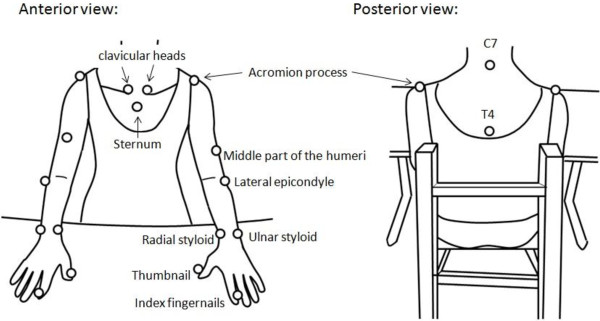
Schematic diagrams (anterior and posterior view) show the upper limb model used for the 3-dimensional markers that were placed on the skin. (○): Retroreflective markers placed on the skin.

Schematic diagrams (anterior and posterior view) show the upper limb model used for the 3-dimensional markers that were placed on the skin. (○): Retroreflective markers placed on the skin.