FIGURE 5.
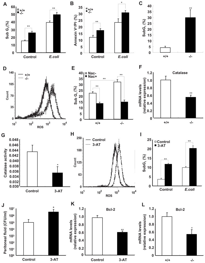
Deficiency of SRC-3 increases apoptosis of peritoneal macrophages due to reduced expression of catalase. A and B, Apoptosis of macrophages from SRC-3−/− mice was significantly higher than that of wild-type mice in the absence and presence of E. coli. The percentage of fragmented DNA (subG1 populations) or Annexin V+/PI− population are indicated in the graphics. Data are the means + SD of four mice per group. ** p<0.01. C, The percentage of apoptotic cells in peritoneal cells from SRC-3−/− mice at 20 h after infection by E. coli is significantly higher than that of wild-type mice. Data are the means + SD of three mice per group. ** p<0.01. D, Deficiency of SRC-3 enhanced the levels of ROS in macrophages. Data shown are representative of three independent experiments. E, Apoptosis of macrophages from both types of mice in the presence of E. coli was decreased after treatment with NAC. Data are the means + SD of three mice per group. ** p<0.01. F, The expression of catalase was determined by real-time PCR. Data are the means + SD of four mice per group. ** p<0.01. G, The catalase activity of wild-type macrophage was inhibited by 3-AT (50 mM). Data are the means + SD of three mice per group. * p<0.05. H, Wild-type macrophages accumulated more ROS after treated with a catalase specific inhibitor 3-AT. Data shown are representative of three independent experiments. I, Apoptosis of wild-type macrophages was increased after treatment with 3-AT. Data are the means + SD of three mice per group, ** p<0.01. J, Pre-treating wild-type mice with 3-AT led to more bacterial loads in peritoneal fluid at 20 h after infection by E. coli. Data are the means + SD of three mice per group, * p<0.05. K, The expression of anti-apoptotic gene Bcl-2 was decreased in wild-type macrophage after treatment with 3-AT. Data are the means + SD of three mice per group, * p<0.05. L, The expression of Bcl-2 was decreased in SRC-3−/− macrophages. Data are the means + SD of four mice per group. * p<0.05.

