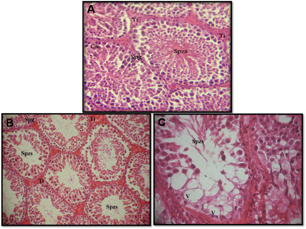
Figure 4.

Testicular sections of control mice which show normal spermatogenesis (part A): Note the normal cell arrangement in the seminiferous tubules. The interstitial spaces also appear normal Ti, interstitium; Sg, spermatogonia; Sd, spermatid; Spzs, spermatozoa; CS, sertoli cell (x 200 H&E). Testicular sections of mice treated with 5 mg/kg/day of DL: Note the atrophic seminiferous tubules (part B) (x400 H&E). Sloughing of germ cells into tubular lumen, V, vacuolization in Sertoli cells and (arrow) multinucleated giant cell (part C) (x1000 H&E).
