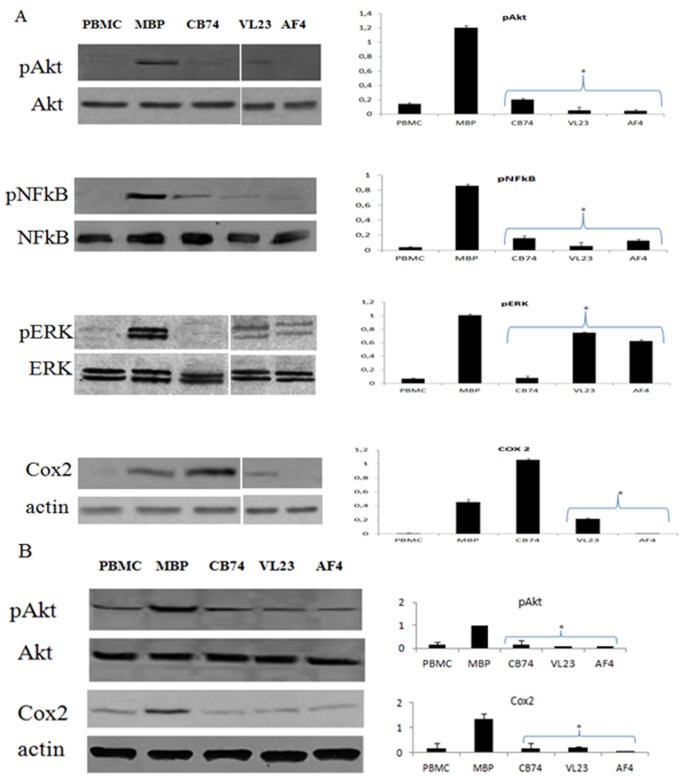
Figure 5. 1,8-naphthyridine, pyridine and quinoline derivatives control Akt, NF-kB, Erk and Cox-2 expression.
MBP activated PBMC were treated with the compounds at the concentration of 10 µM and cultured for 6 days. After the incubation, cell extracts were prepared and protein expression was determined by western blot analysis. In the blots, the bands of un-stimulated cells (PBMC in the figure), MBP activated cells (MBP in the figure) and the treatment with CB74, VL23 and AF4 are showed. The expression of phosphorylated Akt (pAkt) normalized on total Akt (Akt) and Cox-2 normalized on actin is showed in control cells (A) and in patient derived cells (B). The densitometric analysis is also reported in the histograms for both MS patient and control derived cells. In addition, in control cells (A), the protein expression of pNF-kB 65 kDa normalized on total NF-kB 65 kDa and pErk normalized on total Erk is also showed along with their relative densitometric analysis. The blots are representative of four independent experiments and the densitometric analysis reports the mean of the values of all the experiments ± the standard deviation (*p<0,01 significant inhibition calculated with respect to MBP activate PBMC).

