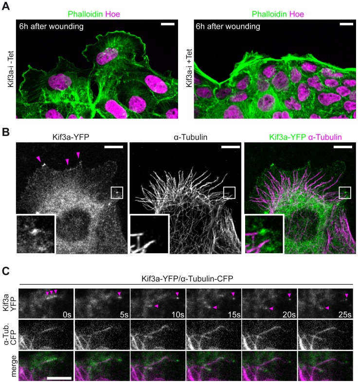
Figure 2. Kif3a associates with MTs at the leading edge.
(A) MDCK.Kif3a-i cells were grown to confluency (48 hours), fixed 6 hours after wounding and stained for actin (phalloidin, green) and the nucleus (Hoechst, magenta). Cell protrusions appear markedly smaller in +Tet conditions. Scale Bars: 10 µm. (B) Migrating MDCK cells expressing Kif3a-YFP (green) were fixed and stained for α-tubulin (magenta). Kif3a-YFP localizes at microtubule tips and cell protrusions (magenta arrows). Scale Bars: 10 µm. (C) MDCK cells stably expressing Kif3a-YFP (green) and α-Tubulin-CFP (magenta) were imaged by dual color TIRF microscopy (Video S2). Kif3a-YFP dynamically co-localizes with growing MTs. Magenta arrows point to Kif3a-YFP accumulations along MTs and MT-tips. Scale Bars: 2 µm.

