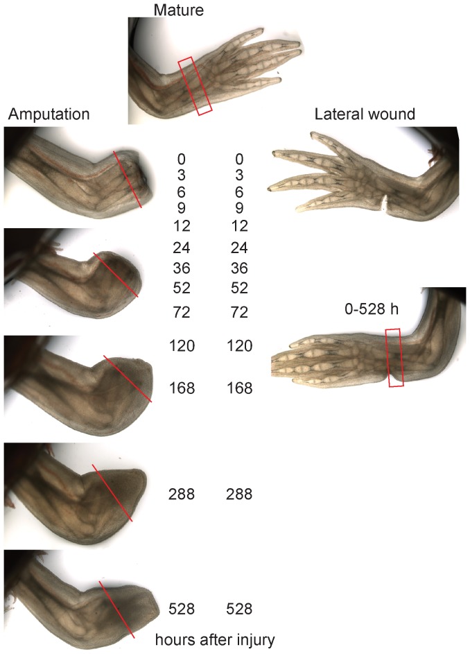
Figure 1. Comparative transcriptome profiling of regeneration versus lateral wounding in the axolotl limb.
Live images of amputation and lateral wound limb samples spanning 0–528 hours (22 days) after injury. The red lines in the amputated series depict the plane approximately 2 mm behind the amputation plane. All tissue distal to the line was collected for the microarray sample. In the mature limb and lateral wound series the collected tissue is depicted by red rectangles.

