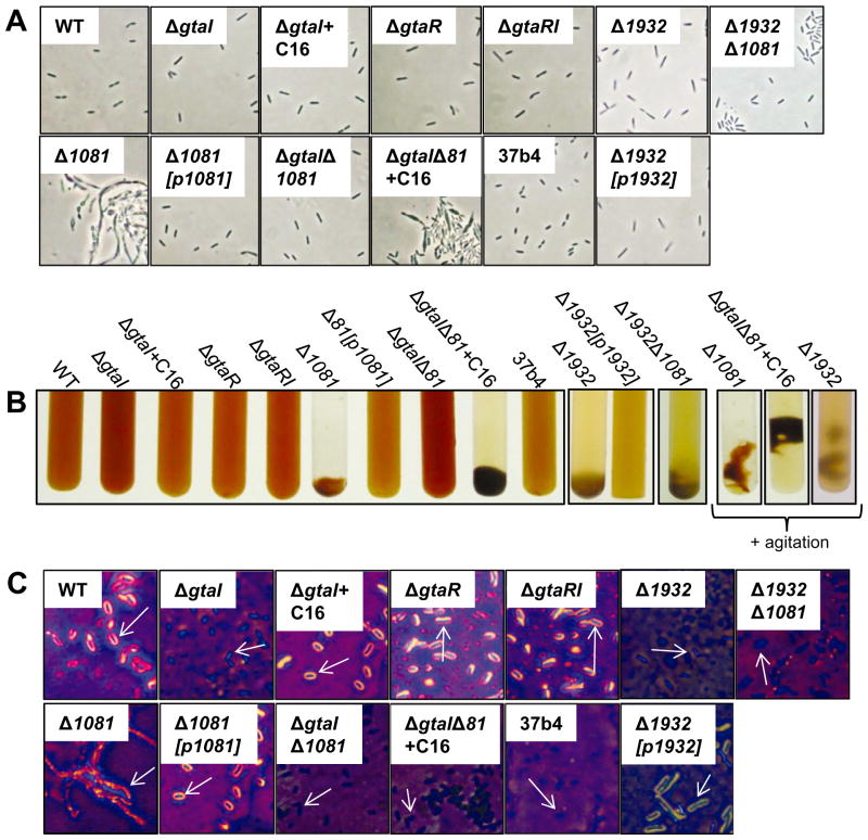
Fig. 5.
Phase contrast microscopy, capsule stain images, and culture macroscopic morphology of WT B10 (WT), ΔgtaI, ΔgtaI + C16-acyl-HSL, ΔgtaR, ΔgtaRI, Δ1081, Δ1081[p1081], ΔgtaI/Δ1081, ΔgtaI/Δ1081 + C16-acyl-HSL, Δ1932, Δ1932[p1932], Δ1932/Δ1081 and WT 37b4 R. capsulatus strains.
A. Phase contrast microscopy images of the strains listed above, at 100× magnification.
B. Images of tubes of phototrophically grown cultures. On the far right, images of agitated cultures of Δ1081, ΔgtaI/Δ1081, and Δ1932 are shown.
C. Capsule stain images of WT, QS and EPS biosynthesis mutant strains.

