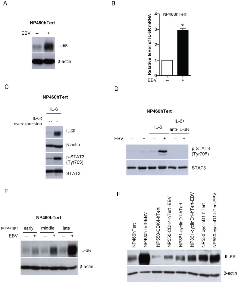
Figure 2. Upregulation of IL-6R in EBV-infected NPE cells is responsible for the enhanced responsiveness to IL-6-induced STAT3 activation.
(A) The expression of IL-6R in EBV-infected and uninfected NP460hTert cells was analyzed by Western blot. Immunoblotting for β-actin was provided as protein loading control. (B) Total RNA was extracted and the expression levels of IL-6R mRNA in NP460hTert and NP460hTert-EBV cells were analyzed by quantitative real time RT-PCR. The mRNA levels of IL-6Rα were normalized to cellular GAPDH mRNA. Data were collected from triplicate separate experiments. * p<0.05. (C) NP460hTert cells were infected with retroviral expression vectors, pBabe-IL-6Rα or pBabe empty vectors. Stably transduced cells were treated with IL-6 at 50 ng/ml for 30 minutes. Cell lysates were prepared and examined for expression of IL-6Rα and p-STAT3 (Tyr 705) by Western blot. Immunoblottings for STAT3 and β-actin are shown as controls for protein loading. (D) NP460hTert and NP460hTert-EBV cells were pre-treated with anti-IL-6R antibody at 5 µg/ml for 30 minutes before IL-6 treatment. The expression of p-STAT3 was analyzed by Western blot. Immunoblotting for total levels of STAT3 protein is shown as controls for protein loading. (E) After EBV infection, the EBV-infected NP460hTert cells and its uninfected parental cells were continuously subcultured. Cells lysates were prepared from the cells that had been passaged for 56, 99 and 133 times (designated as early, middle and late passage). The expression of IL-6R was analyzed by Western blotting. Immunoblotting for β-actin was included as control for protein loading. (F) The levels of IL-6R expression in cells were detected by Western blot in several paired uninfected and EBV-infected cell lines, including EBV-infected and non-infected pairs of NP460hTert, NP550-CDK4-hTert, NP361-cyclinD1-hTert, NP550-cyclinD1-hTert. β-actin expressions were shown as protein loading control.

