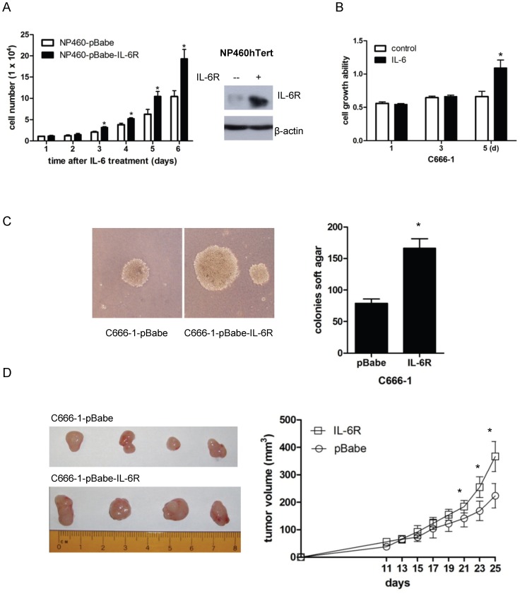
Figure 5. IL-6R overexpression promotes growth of immortalized NPE cells in vitro and tumorigenic properties of NPC cells in vivo.
(A) pBabe control or pBabe-IL-6Rα expressing NP460hTert cells (1×10⁴ per sample) were cultured in the presence of IL-6. Cells were counted at the indicated time points. Data represent the mean of triple independent experiments ± SD. * p<0.05. The right panel shows the Western blot analysis of the expression of IL-6R in the NP460-pBabe-IL-6R cells and the control NP460-pBabe cells. (B) Effect of IL-6 on the growth of C666-1 cells. C666-1 cells were treated with or without IL-6 at 50 ng/ml. The MTT assay was conducted on the indicated time intervals. Each datum point represents the average of triplicate experiments. * p<0.05. (C) C666-1 cells were transduced with pBabe-IL-6R or pBabe by retroviral gene transfer. Vector- and IL-6R-expressing C666-1 cells were examined for colony formation in soft agar. Representative images of the soft agar colonies were shown. The number of colonies presented is mean ± SD from triplicate results. The difference in the number of soft agar colonies between pBabe- and IL-6R-expressing C666-1 cells is statistically significant (* p<0.05). (D) pBabe vector control- and IL-6R-expressing C666-1 cells (1×10⁶ per mouse) were subcutaneously injected into nude mice. Tumor size was measured at the indicated time points. Each time point of growth curve was calculated as the mean ± SD of five tumors per experimental group. The differences in the growth rate in xenografts established from control- and IL-6R-expressing C666-1 cells were statistically significant after injecting the cells for 21 days (* p<0.05).

