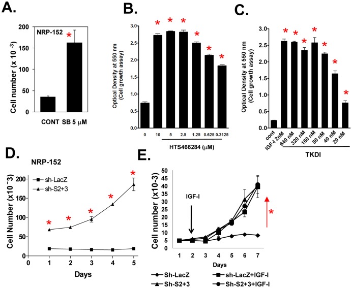
Figure 6. IGF-I enhances cell growth by suppressing TGF-β autocrine signaling.
(A–C) TGF-β receptor kinase inhibitors stimulate the growth of NRP-152 cells. NRP-152 cells plated were treated with 5 µM of SB431542 (SB) (A) or with various concentrations of HTS466284 or TKDI and cell growth was measured 6 days later by counting cell number (A) or by crystal violet staining of fixed cells (B,C). (D) Growth of NRP-152-Sh-Smad2+3 cells versus NRP-152-Sh-LacZ cells in GM3 medium. (E) NRP-152-sh-LacZ and NRP-152-sh-Smad2+3 cell lines were incubated in the presence or absence of LR3-IGF-I (2 nM) for 5 days and cell growth was monitored daily for 5 days (D,E). Percent of growth inhibition by rapamycin in NRP-152-sh-LacZ and NRP-152-sh-Smad2+3 cell lines. Cell numbers were measured using Coulter Electronics Counter. Data shown are the average of triplicate determinations ± S.E. (*p<0.01). Statistical significance (*p<0.01) was assessed by two-way Anova analysis of variance.

