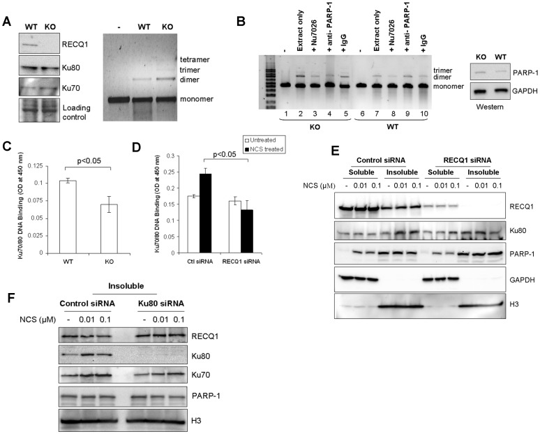
Figure 6. RECQ1-null cells promote end-joining but show reduced Ku-DNA binding activity.
A. Level of in vitro end-joining activity is comparable in WT and RECQ1 KO MEFs. Cell free extracts prepared from non-transformed RECQ1 WT or KO MEFs were used in end-joining reaction containing EcoRI-linearized pUC19 DNA as substrate. Linear substrate DNA is indicated as monomer, and the end-joined products corresponding to dimer, trimer and tetramer are indicated (right panel). Western blot showed no detectable difference in Ku protein level in WT and KO cell extracts (left panel). GAPDH serves as loading control. B. Presence of PARP-1 antibody interferes with RECQ1 KO cell free extract mediated end-joining. In addition to standard reactions, in vitro end-joining reactions were performed with WT or KO cell extracts in the presence of a DNA-PKcs inhibitor Nu7026 (1.2 µM) or a specific anti-PARP-1 antibody (3 µg). IgG (3 µg) was included as unrelated antibody in a control reaction. Linear substrate DNA is indicated as monomer, and the end-joined products corresponding to dimer and trimer are indicated. Western blot showed no detectable difference in PARP-1 protein level in WT and KO cell extracts (left panel). GAPDH serves as loading control. C. Ku70/80 DNA binding assay performed by using Active Motif kit shows diminished DNA binding in RECQ1 KO extract as compared to WT extract (p<0.05). The results shown are the average of at least three independent experiments, with SD indicated by error bars. D. DNA binding analysis for Ku70/80 in extracts prepared from control or RECQ1-depleted HeLa cells. Nuclear extracts prepared from control or RECQ1 siRNA transfected cells either untreated or treated with NCS (0.01 µM, 3 h) were used to perform Ku70/80 DNA binding assay (Active Motif). Following NCS treatment, RECQ1 siRNA transfected cells showed significantly reduced Ku70/80 DNA binding activity as compared to control siRNA transfected cells (p<0.05). The results shown are average of at least three independent experiments, with SD indicated by error bars. E. RECQ1-depleted HeLa cells show reduced chromatin bound Ku following NCS treatment. Ku80 and PARP-1 was detected by Western blot in the soluble and insoluble fractions prepared from control or RECQ1 siRNA transfected cells that were either untreated or treated with indicated dose of NCS for 3 h. GAPDH serves as cytoplasmic marker and H3 serves as marker for chromatin enriched fractions. F. RECQ1 protein in the chromatin enriched fraction of control or Ku80-depleted cells. Cells were untreated or treated with indicated dose of NCS for 3 h, followed by biochemical fractionation and the chromatin containing insoluble fractions were examined by Western blotting. H3 serves as marker for chromatin enriched fractions.

