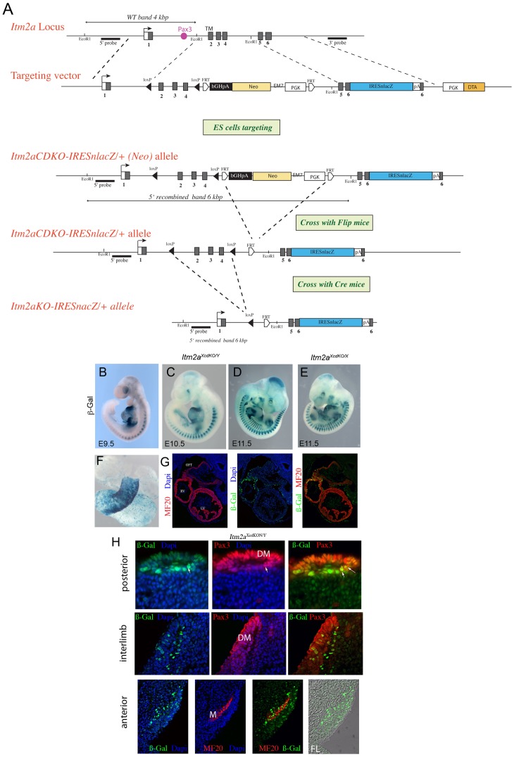
Figure 2. Generation of an Itm2a conditional reporter allele.
A. Schematic diagram of the Itm2a locus and targeting construct. The construct contains loxP sites inserted into exon 1 and 4. A PGK-Neo-pA (Neo) selection marker is flanked by FRT sites and inserted into exon 4. An IRES-nLacZ cassette has been inserted into the 3′ UTR (exon 6). A counter-selection cassette encoding the A subunit of Diphtheria Toxin was inserted at the 5′ end of the targeting vector. Schematic diagrams of the Itm2acdKO-IRESnLacZ (Neo) and Itm2aKO-IRESnLacZ (Neo) alleles are also shown. Probes and restriction enzymes are indicated, with the size of the resulting wild-type and recombined restriction fragments. Pax3 sites located in Itm2a intron1 are represented by a pink box. B–D, X-Gal stained Itm2aXcdKO/Y embryos at E9.5 (B), E10.5 (C) and E11.5 (D). E, An X-Gal stained Itm2aXcdKO/X embryo at E11.5. Note the chimeric expression of the reporter, due to random X inactivation. F, An isolated X-Gal stained heart of an Itm2aXcdKO/Y embryo at E9.5. G, Immunohistochemistry on transverse sections through the heart region of an E9.5 Itm2aXcdKO/Y embryo using antibodies recognizing striated muscle myosin MF20 (red), β-Gal (green). Dapi staining of nuclei is in blue. The right hand panel shows a merged image with both antibodies. OFT, outflow tract; RV, right ventricle; LV, left ventricle. H, Immunohistochemistry on transverse sections through immature somites of an Itm2aXcdKO/Y embryo at E10.5, in the more caudal region (top), in the interlimb region (middle) and just anterior to the forelimb (bottom), using antibodies recognizing Pax3 or striated muscle myosin (MF20) (red) as indicated on the Figure, and β-Gal (green). Dapi staining of nuclei is in blue. Merged images are shown on the right. In the bottom series, the right hand panel shows phase contrast with β-Gal on a somite just anterior to the forelimb bud. Dermomyotome (DM), Myotome (M), Forelimb (FL). Arrows point to cells expressing Pax3 and ßGal (Itm2a).

