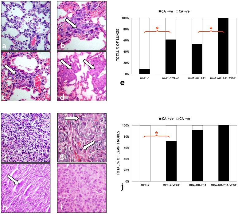
Figure 7. Histological validation of the transformation of non-invasive MCF-7 tumors to the metastatic phenotype following VEGF overexpression.
Representative (40×) images of H&E stained lung sections from: (a) MCF-7, (b) MCF-7-VEGF, (c) MDA-MB-231 and (d) MDA-MB-231-VEGF tumor bearing animals. Cancer cells are indicated by solid arrows in each panel. (e) Comparison of the number of animals with cancer positive (CA +ve) and cancer negative (CA -ve) lungs for each type of breast cancer xenograft. Representative (40×) images of H&E stained lymph node sections from: (f) MCF-7, (g) MCF-7-VEGF, (h) MDA-MB-231 and (i) MDA-MB-231-VEGF tumor bearing animals. Cancer cells are indicated by solid arrows in each panel. (j) Comparison of the number of animals with cancer positive (CA +ve) and cancer negative (CA -ve) lymph nodes for each type of breast cancer xenograft.

