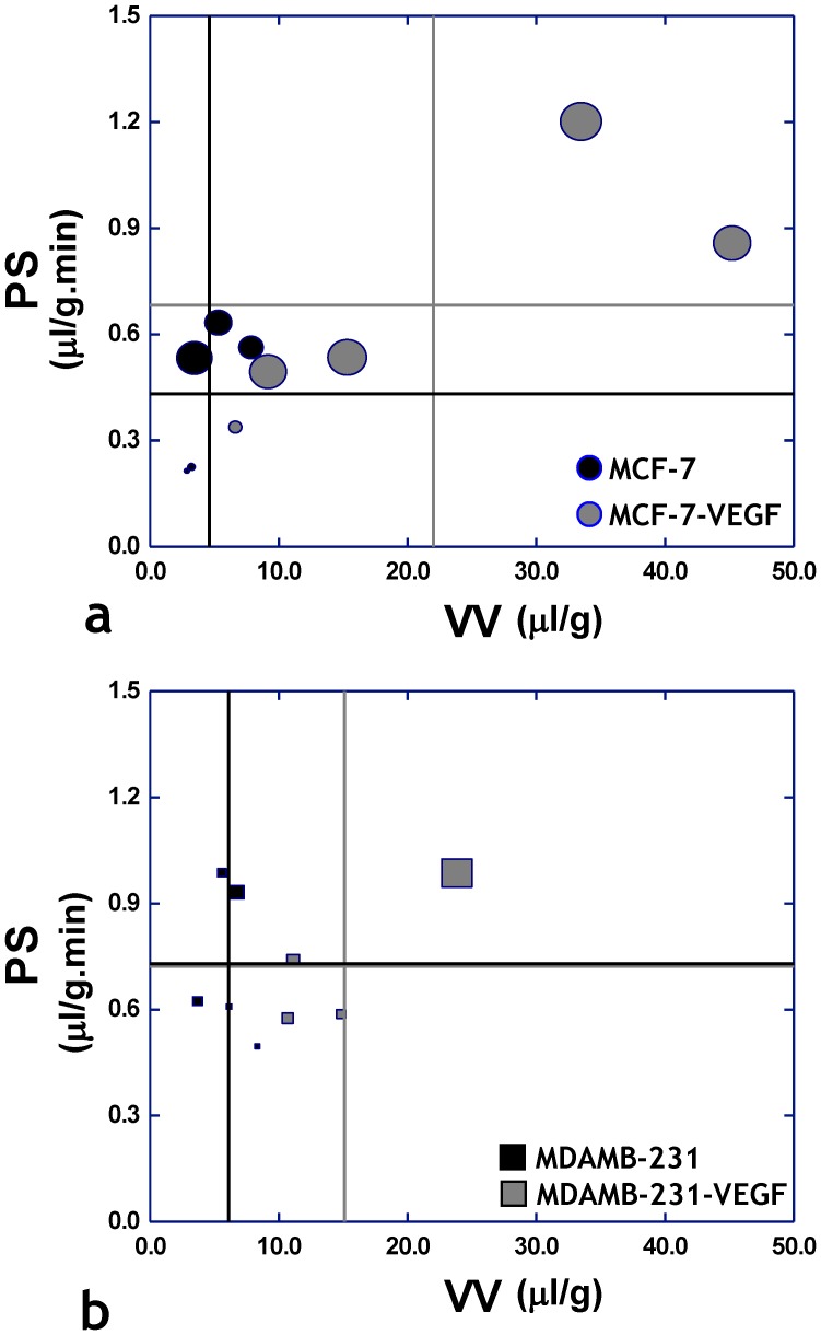
Figure 8. “MRI phenotyping” reveals progression of the non-invasive MCF-7 human breast cancer model to the metastatic phenotype is driven by VEGF overexpression.
Scatter plots of permeability-surface area (PS) product versus vascular volume (VV) for each tumor type, in which each symbol is scaled according to the volume of extravascular fluid drained. The vertical lines represent the mean value of VV for each tumor type, while the horizontal lines represent the mean value of PS for each tumor type. (a) Scatter plot of parameters measured in vivo from MCF-7 (n = 5) and MCF-7-VEGF (n = 5) tumors reveal a trend in the MCF-7-VEGF group towards elevated PS and VV values (gray crosshairs) in conjunction with greater extravascular fluid drainage (larger symbol size) – which collectively indicate the progression of the original MCF-7 phenotype to a metastatic one. In contrast, (b) a scatter plot of parameters measured in vivo from MDA-MB-231 (n = 5) and MDA-MB-231-VEGF (n = 4) tumors does not show this trend, but shows the elevation of VV in the MDA-MB-231-VEGF tumors.

