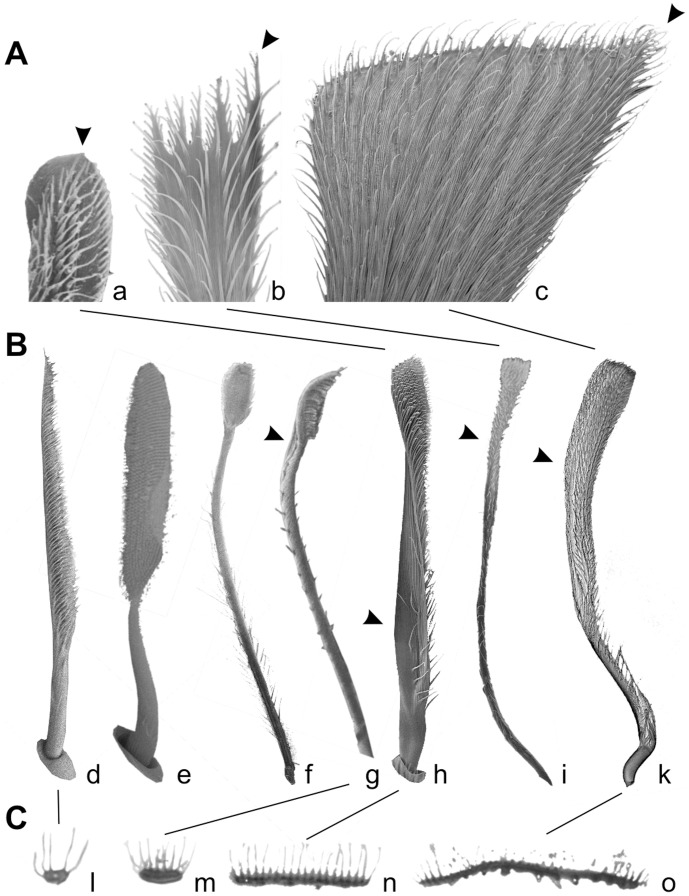
Figure 4. Electron microscopy of isolated setae, different scales.
A. SEM micrographs of distal tips of claw tuft setae, rear view. Arrowheads indicate the remaining tapered tip of the expanded setae. a. Adhesive seta type IIb in Micaria formicaria (Gnaphosidae). b. Adhesive seta type III in Clubiona pallidula (Clubionidae). c. Large adhesive seta type IIa in Anyphaena accentuata (Anyphaenidae). B. SEM micrographs of setae, lateral view. Arrowheads indicate the twisted lamella shaft occurring in claw tuft setae. d. Frictional seta type II in Xysticus lanio (Thomisidae) ventral tarsus. e. Scopula seta of type IIb in Clubiona lutescens (Clubionidae) prolateral tarsus. f. Scopula seta of type IIb in Palpimanus gibbulus (Palpimanidae) prolateral metatarsus. g. Brush like claw tuft seta of type Ia in Homalonychus selenopoides (Homalonychidae), a presumably primitive character. h. Claw tuft seta of type IIb in Euophrys frontalis (Salticidae). i. Claw tuft seta of type III in Clubiona pallidula. k. Claw tuft seta of type IIa in Anyphaena accentuata. C. TEM micrographs of sections of the distal part of tarsal setae. l. Frictional seta type II in Nops largus (Caponiidae). m. Adhesive seta type Ia in Xysticus cristatus (Thomisidae). n. Adhesive seta type IIb in Evarcha arcuata (Salticidae). o. Adhesive seta type IIa in Anyphaena accentuata.

