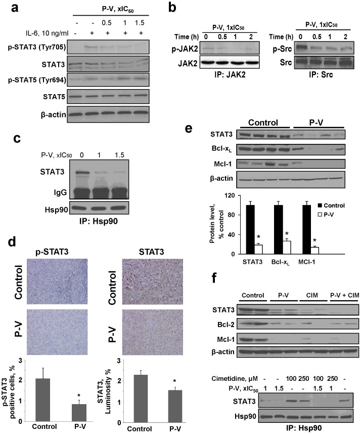
Figure 2. P-V inhibits STAT3 signaling in vitro and in vivo.
(A) Immunoblots of STAT3, phosphorylated STAT3 (p-STAT3), STAT5 and p-STAT5 from BxPC-3 cells treated with P-V for 1 h and stimulated with IL-6 for 30 min. (B) JAK2 and Src were immunoprecipitated from control and P-V-treated MIA PaCa-2 cells and the precipitates were immunoblotted. (C) Hsp90 was immunoprecipitated from control and P-V-treated BxPC-3 cells and the precipitate was immunoblotted for STAT3. IgG = Loading control. (D) Immunostaining for STAT3 and phosphorylated STAT3 (p-STAT3) expression on tissue sections of MIA PaCa-2 orthotopic tumors from control and P-V-treated mice (x40). Results are expressed as percent of positive cells for p-STAT3 and luminosity index per 40×field for STAT3. Values are mean±SEM (n = 7); *p<0.05 vs. control. (E) Protein lysates from BxPC-3 xenografts were immunoblotted for STAT3, Bcl-xL and Mcl-1 proteins. Each lane represents a different tumor sample. Loading control: β-actin. Bands were quantified and results expressed as percent control for each protein. Values are mean±SEM (n = 7); *p<0.01 vs. control. (F) (Upper) Cimetidine (CIM) enhances P-V's inhibitory effect on STAT3 in vivo. Protein lysates from MIA PaCa-2 xenografts were analyzed for STAT3, Bcl-2 and Mcl-1 proteins by immunoblotting. Each lane represents a different tumor sample. Loading control: β-actin. (Lower) CIM enhances P-V's disruption of the STAT3-Hsp90 association. Hsp90 was immunoprecipitated from MIA PaCa-2 cells treated with or without P-V, CIM or both. After immunoprecipitation, immunoblotting for STAT3 was performed.

