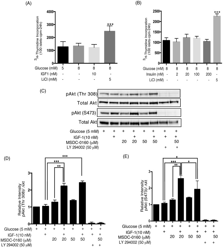
Figure 1. Human islets display insulin signaling pathway resistance.
(A, B) Human islets do not increase DNA synthesis in response to nutrients or insulin/IGF-1 in vitro. Human islets (100) were cultured for 4 days in cCMRL, 8 mM glucose+MSDC-0160, insulin (A) or IGF-1 (B) and LiCl as indicated. 3H-thymidine was added to each dish 24 h before the end of the 4-day period. Data are the means±SEM from n = 3, with duplicate samples in the experiment. Data analyzed using one-way ANOVA followed by Newman-Keuls Post-hoc test. (C–E) PPARγ sparing TZD, MSDC-0160, restores the insulin-signaling pathway. Human islets were cultured for 90 min in cCMRL, 5 mM glucose with or without the concentrations of MSDC-0160, IGF-1, LY 294002 as indicated. Cell extracts were prepared and samples were processed for Western blotting and quantitated by densitometry. Following detection of the phosphorylated proteins, the Western blots were reprobed with antibodies against their respective endogenous control (Total Akt). In each figure, the upper panel (Fig. C) shows representative Western blots and the lower panels (Fig. D&E) shows mean values after quantitation of the data. Data analyzed using one-way ANOVA followed by Newman-Keuls test. Data are means±SEM of n = 3 (D) or n = 4 (E) experiments.

