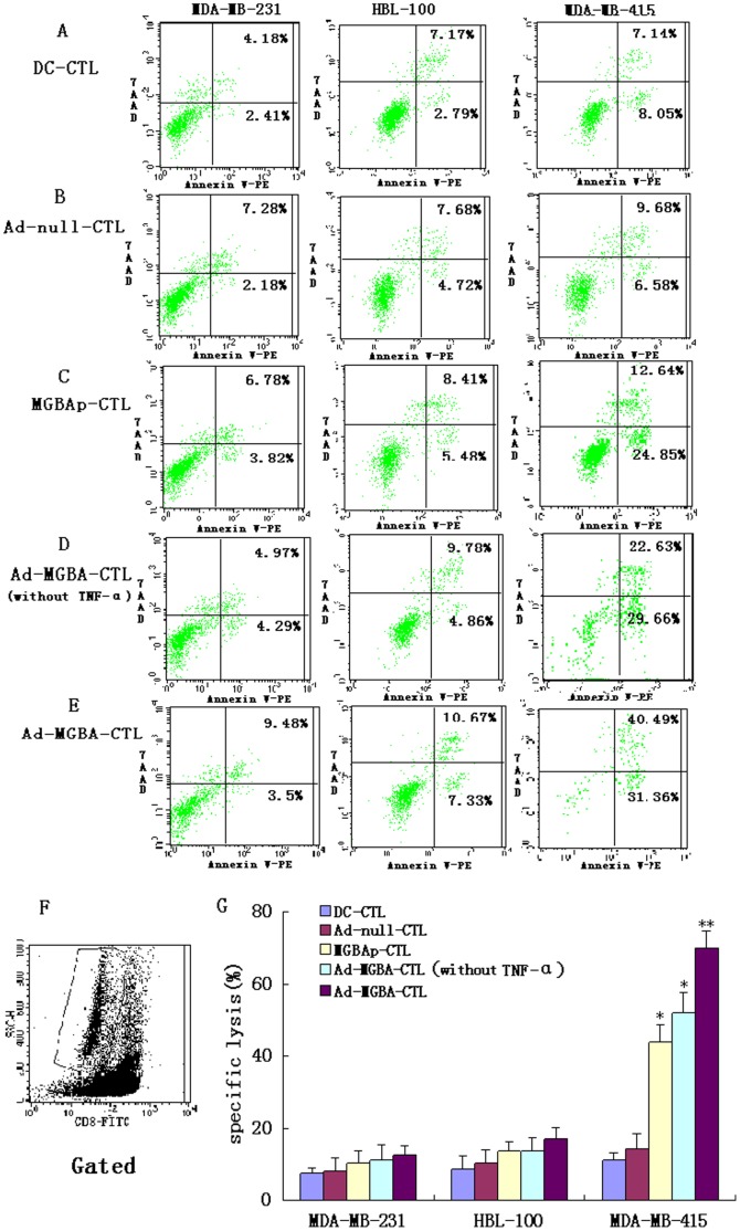
Figure 6. The cytotoxic effects of MGBA specifically stimulated CD8+CTLs on breast cancer cells.
Four CD8+CTLs co-cultured with different DCs treated with TNF-α (i.e., DC-CTL, Ad-null-CTL, MGBAp-CTL, and Ad-MGBA-CTL) and CD8+CTLs co-cultured with DCs transfected only with Ad-MGBA (i.e., Ad-MGBA-CTL without TNF-α ) were added into breast cancer cell cultures at an E:T ratio of 20∶1. 12 h later, apoptosis rates of these breast cancer cells were analyzed by FACS. (A) DC-CD8+CTLs; (B) Ad-null infected DC-CD8+CTLs; (C) MGBAp-CD8+CTL; (D) Ad-MGBA infected DC-CD8+CTLs (without TNF-α); (E) Ad-MGBA infected DC-CD8+CTLs; (F) Non-FITC-CD8-conjugated breast cancer cells in co-culture cells gated; (G) Histogram summarizing the data from six independent experiments from two volunteers. The apoptosis rate in HLA-A33+/MGBA+ MDA-MB 415 cells induced by Ad-MGBA-CD8+CTL was the highest among these five CTLs (p<0.01). * indicates that this group has statistically significant differences compared to the others (p<0.05). ** indicates that this group has statistically significant differences compared to the others (p<0.01).

