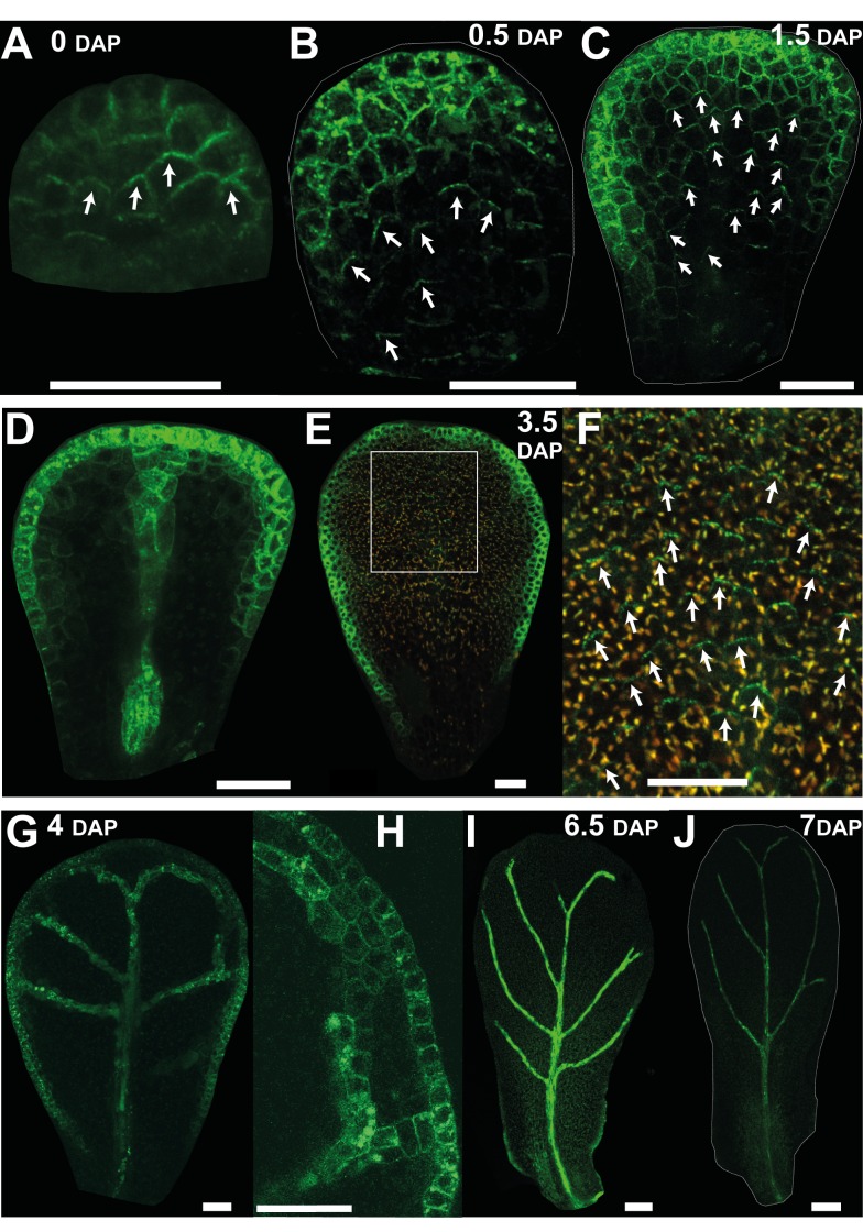
Figure 6. Epidermal expression pattern of the auxin efflux carrier PIN1 in petals supports a divergent polarity field.
(A) At 0 DAP, epidermal PIN1::GFP is preferentially localised to the distal end of cells in the plane of the petal. White arrows on cells point towards the centre of the distal PIN1 expression. (B, C) At 0.5 and 1.5 DAP, PIN1 polarity in the epidermis points divergently toward the distal end of the tissue. Strong expression is also seen at or near the petal margin but without clear polar localisation. (D) Deeper section of (C) showing expression in the provascular tissue. (E) At 3.5 DAP, PIN expression in the main plain of the epidermis is weak. (F) Enlargement of the white box in (E) showing PIN1 signal at the distal end of the epidermal cells. PIN1 expression is lower so the GFP channel is shown merged with the red channel (corresponding to the emission of chlorophyll autofluorescence) to facilitate visualisation. (G) At 4 DAP, PIN1 signal is no longer detected in the main plane of the petal but can be observed in the petal margin and vascular tissue. (H) Close up of (G). (I) At 6.5 DAP, expression at the distal margin has disappeared. (J) Little signal is observed from 7 DAP onwards. Width of petals: (A–C) 30, 45, and 75; (E) 146; (G) 170; (I) 343; (J) 425 µm. Scale bar, 20 µm (A–H), 50 µm (I), and 80 µm (J).

