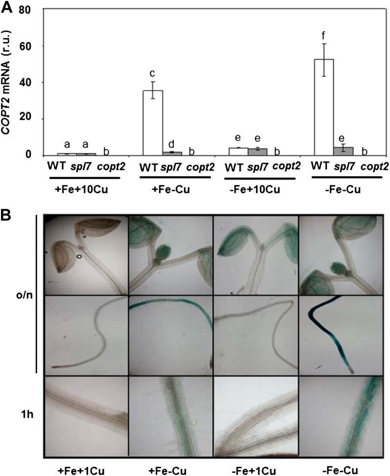
Figure 3.
COPT2 expression under Cu and Fe deficiencies. A, COPT2 expression analysis by qPCR in wild-type (WT; white bars), spl7 (gray bars), and copt2-1 (copt2; dark gray bars) seedlings. Total RNA from 7-d-old seedlings grown under the control (+Fe+Cu; supplemented with 10 μm CuSO4), Cu deficiency (+Fe−Cu), Fe deficiency (−Fe+Cu; supplemented with 10 μm CuSO4), or Fe and Cu deficiency (−Fe−Cu) conditions was isolated and retrotranscribed to complementary DNA. UBQ10 gene expression was used as a loading control. Values are means ± sd of three biological replicates. r.u., Relative units. Different letters above the bars represent significant differences among all the means (P < 0.05). B, GUS staining in 7-d-old seedlings from the PCOPT2:GUS transgenic lines grown under control (+Fe+Cu; supplemented with 1 μm CuSO4), Cu deficiency (+Fe−Cu), Fe deficiency (−Fe+Cu; supplemented with 1 μm CuSO4), or Fe and Cu deficiency (−Fe−Cu) conditions at different incubation times at 37°C (overnight [o/n] and 1 h). [See online article for color version of this figure.]

