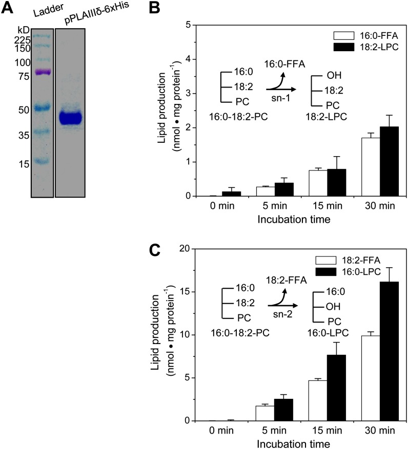
Figure 5.
pPLAIIIδ was purified and hydrolyzes PC at the sn-1 and sn-2 positions. A, Coomassie blue staining of an 8% SDS-PAGE gel loaded with affinity-purified pPLAIIIδ-6×His from E. coli. B, Production of 16:0-FFA and 18:2-LPC from hydrolysis of 16:0-18:2 PC at the sn-1 position (inset). Values are means ± se (n = 3 separate samples). C, Production of 18:2-FFA and 16:0-LPC from hydrolysis of 16:0-18:2 PC at the sn-2 position (inset). Values are means ± se (n = 3). [See online article for color version of this figure.]

