Figure 4.
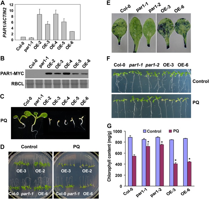
Overexpression of PAR1 confers hypersensitivity to paraquat. A, Analysis of PAR1 overexpression in Col-0, par1-1, and Super:PAR1-MYC (OE lines) using qRT-PCR. RNA prepared from 2-week-old seedlings was used for this assay, and the means of three replicates ± sd are shown. Similar results were obtained in three independent experiments. B, Immunoblot analysis of PAR1 protein in the plants described in A using an anti-MYC antibody. Equal loading was verified by Coomassie blue staining. RBCL, Rubisco large subunit. C, Ten-day-old seedlings with the indicated genotypes germinated and grown in the presence of 0.5 μm paraquat (PQ). D, Phenotypes of Col-0, par1-1, and three independent overexpression seedlings (5 d old) grown on MS medium were transferred to MS medium supplemented with water (control) or 1 μm paraquat for an additional 7 d. E, Paraquat-induced cell death in leaves of wild-type (Col-0), par1, and PAR1-OE plants (4 weeks old) treated with water or 5 μm paraquat for 24 h by spraying. Leaves were then detached from treated plants and stained with Evans blue. F, Phenotypes of wild-type (Col-0), par1, and PAR1-OE seedlings (10 d old) transferred onto MS medium supplemented with 10 μm paraquat for 48 h. G, Chlorophyll content in the seedlings shown in F. Asterisks indicate P < 0.05 (Student’s t test) when compared with the paraquat-treated Col-0. [See online article for color version of this figure.]

