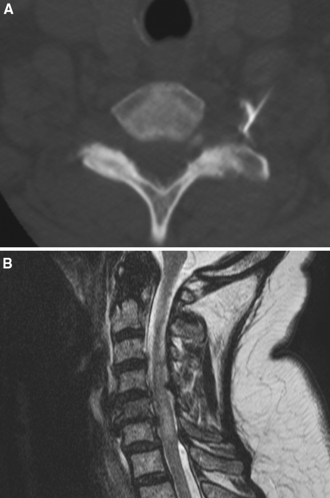
Fig. 1.

Imaging studies of a 71 year old female (Case 1). a C7/T1 foraminal injection on the left side. Needle position outside the intervertebral foramen. A tentative injection of contrast media demonstrated extraforaminal contrast distribution. b T2-weighted sagittal MR image obtained 1 day after injection demonstrating ischemic myelopathy at the C5–C7 levels
