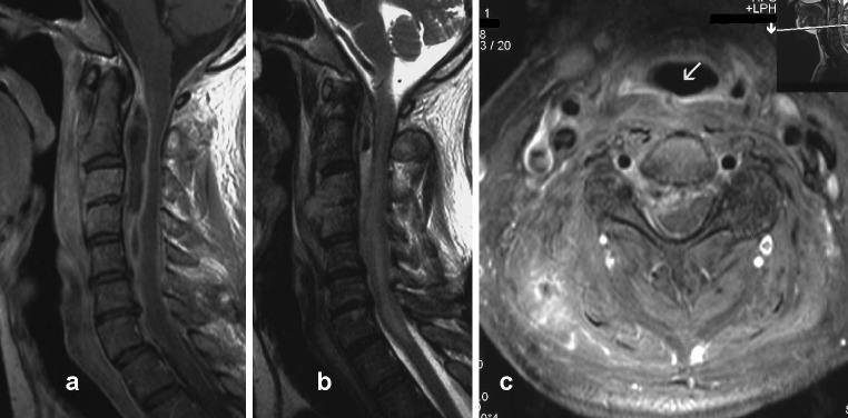
Fig. 2.

Post-contrast sagittal (a) and axial (c) T1-weighted, and sagittal T2-weighted (b) images of the cervical spine. Images illustrate a ventral-lateral right epidural collection that is hyperintense in T2 W and hypointense in T1 W sequences with an enhancing rim and small amount of gas in its rostral part. Cervical spinal cord is diffusely hyperintense in T2 W images. The C3–C4 disc space is obliterated and vertebral bodies of C3 and C4 are hyperintense in T2 W with well enhancement in T1 W sequences, reflecting discitis and osteomyelitis. Infection of retropharyngeal and paravertebral soft tissues is evident in all figures. Fistulous channel with good rim enhancement (arrow) at the C3–4 level is visualized in axial image
