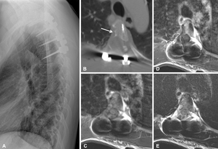
Fig. 4.
Postoperative imaging. Lateral view thoracic spine radiography with instrumentation at levels T2–T5 (a). Axial computed tomography image of the T4 vertebra shows the right-sided costotransversectomy, resection of the right pedicle and posterior elements, and the corticalized scalloping of the vertebral body (arrow) (b). Magnetic resonance imaging (MRI) demonstrates complete resection of the tumor without residual contrast enhancement, and re-expansion of the thecal sac and spinal cord on T1-weighed axial image (T1WI) (c), following contrast administration (d), and on T2WI image (e)

