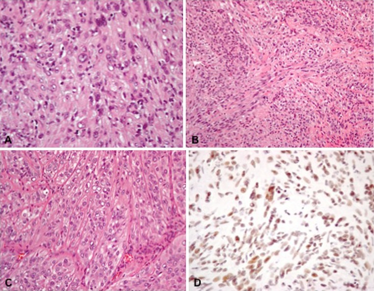
Fig. 5.
Histopathological microphotographs of the tumor. a Epithelioid cells with vacuolated nuclei and prominent nucleoli (hematoxylin eosin, original magnification ×400). b Spindle cell component of the tumor with poorly defined fascicular pattern (hematoxylin eosin, original magnification ×200). c Prominent capillaries in the epithelioid part of the tumor (hematoxylin eosin, original magnification ×200). d immunohistochemistry for integrase interactor 1 protein (INI-1, original magnification ×400)

