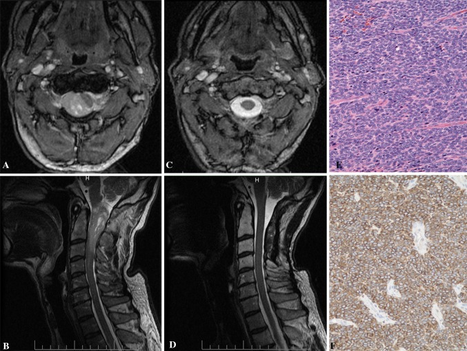
Fig. 2.
a, b Preoperative T1 sagittal and axial MRI with contrast showed a enhancing lesion at the C1–3 levels with severe compression of the spinal cord. c, d Postoperative T1 sagittal and axial MRI with contrast demonstrating resection of the tumor. e, f H&E stained sections disclosed tightly packed oval nuclei forming fascicles (e). Scattered tumor cells were positive for CD34 (f). Original magnification ×200

