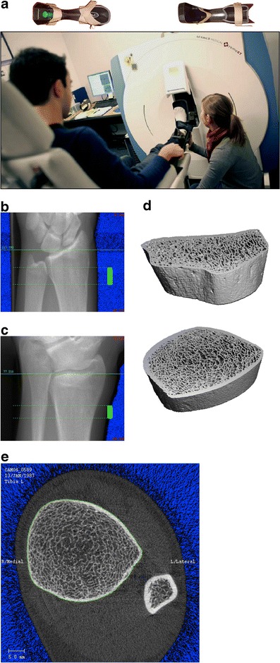
Fig. 2.

a, A typical setup of HR-pQCT for patient measurements. At top, the casts used for securing the forearm and lower leg is shown. b, c, and d, The procedure for HR-pQCT analysis requires a ‘scout view’ x-ray of the radius or tibia (left) so that the operator can select the region of interest for scanning (solid green bar). Subsequently, three-dimensional data can be obtained in the scan region (right). e, A typical section from HR-pQCT showing the ultradistal tibia and fibula. The green line on the periosteal surface of the tibia is used to extract the bone of interest for subsequent three-dimensional analysis
