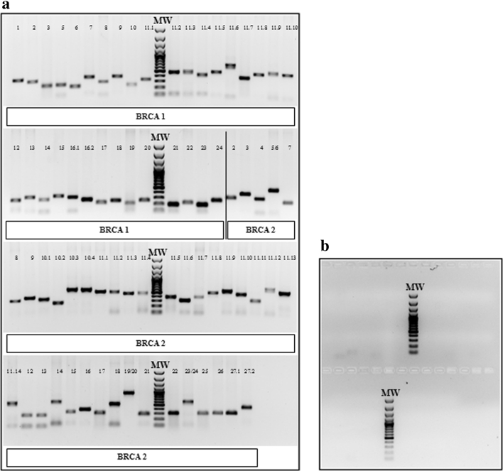
Fig. 1.
PCR analysis of BRCA1/2 coding regions. a 1 % agarose gel electrophoresis of the PCR products to verify the correct amplification of the BRCA1/2 coding regions. MW: molecular weight marker. Each lane corresponds to the amplicon relative to the indicated BRCA1 or BRCA2 exon. In particular, for BRCA1, we divided exon 11 and exon 16 in ten and two amplicons, respectively (11.1–11.10 and 16.1–16.2); for BRCA2 we divided exon 10, exon 11, and exon 27 in three, fourteen, and two amplicons, respectively (10.1–10.3; 11.1–11.14, and 27.1–27.2). For BRCA2 only, exon 5 and exon 6, exon 19 and exon 20, exon 23 and exon 24 were amplified within the same amplicon because they were sufficiently short. b 1 % agarose gel electrophoresis to control the specificity of amplification. No bands were detected in the mix of negative controls. MW: molecular weight marker

