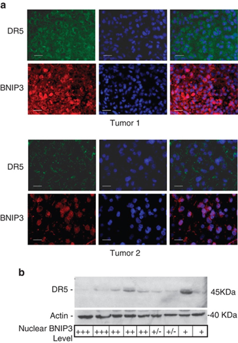
Figure 5.
Primary GBM tumors that have high nuclear BNIP3 levels have low DR5 expression (a) Paraffin sections of representative primary GBM tumors sections were immunostained with antibodies against BNIP3 (red) and DR5 (green). DNA was stained with DAPI (blue), and the slides were analyzed on an Olympus fluorescence microscope. Scale bar represents 20 μM. (b) Representative frozen GBM tumor tissues were lysed to extract total protein, and analyzed by western blot for DR5 expression. The blots were stripped and reprobed with actin antibodies for loading control. The grading for nuclear BNIP3 levels (determined by immunofluorescence) is indicated for each tumor. Nuclear staining was graded as: +++ for high nuclear staining, ++ for moderate nuclear staining,+for low nuclear staining, and +/− for undetectable nuclear staining. Three independent experiments were quantified by densitometry with Quantity One (Bio-Rad) and averaged to obtain DR5 expression relative to actin loading controls

