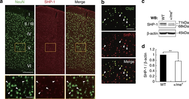
Figure 1.
SHP-1 is expressed in cortical neurons. (a) NeuN (green) and SHP-1 (red) staining in the adult cerebral cortex (upper panels) and layer V of cerebral cortex (lower panels). Arrowheads indicate the expression of SHP-1 in the layer V neurons. Scale bars: upper, 200 μm: lower, 50 μm. (b) Ctip2 (green) and SHP-1 (red) staining in the adult motor cortex. Arrowheads indicate the expression of SHP-1 in the Ctip2-positive neurons. Scale bar, 50 μm. (c) SHP-1 expression in wild-type and +/mev mice. The expression level of SHP-1 was examined by western blot analysis. (d) SHP-1 signal intensity was quantified by densitometry and normalized to β-actin. Data are presented as mean±S.E.M (n=5, each group). **P<0.01, Student's t-test

