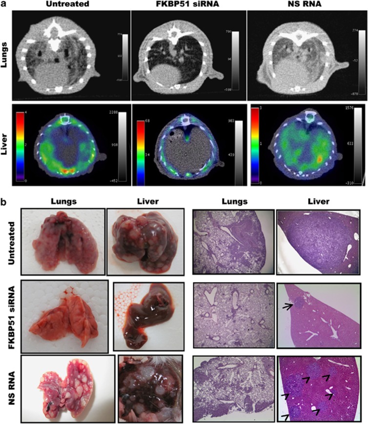
Figure 4.
FKBP51 siRNA counteracts metastasis formation. (a upper) CT images of lungs axial view. In the untreated and NS RNA-treated mouse, marked thickening of the lung interstitium and peribronchial tissue; in the FKBP51 siRNA-treated mouse, lung parenchyma shows no significant abnormality. (a lower) ([18]F-FDG PET/CT images of liver, axial view. Increased FDG uptake throughout untreated or NS RNA-treated liver. No FDG uptake in right or left FKBP51 siRNA-treated liver lobes. (b, left) Representative lungs and liver excised from mice. Organs from untreated or NS RNA-treated mice were completely substituted by nodules. Organs from FKBP51 siRNA-treated mice were apparently not altered morphologically. (b right) Haematoxylin and eosin (H/E) staining of lung and liver biopsies. Lung specimens (H/E; × 50): large metastatic melanoma nodules extensively involve the lung specimen from untreated mouse and NS RNA mouse; in lung biopsy from a FKBP51 siRNA-treated mice, metastatic cells form small peribronchial and perivascular aggregates. Liver specimens (H/E; × 25): the metastasis extensively substitutes the liver tissue in untreated mouse; only a single small subcapsular nodule is visible in liver biopsy from FKBP51 siRNA-treated mouse; NS RNA mouse liver is completely substituted by multiple nodules

