Figure 3.
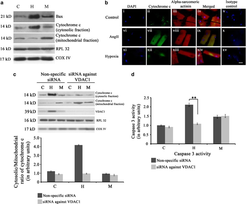
Activation of mitochondrial apoptotic machinery during hypertrophy. (a) Western blot analyses showing significant increase in the expression of Bax and cytochrome c in hypertrophy (H) compared with either MI (M) or sham control (C). RPL32 was used as loading control for cytosolic proteins whereas COX IV was used as loading control for mitochondrial proteins. Data is representative of three independent experiments. (b) Immunofluorescence study showing upregulation of mitochondrial apoptotic marker cytochrome c in hypertrophic cardiomyocytes. Adult cardiomyocytes were stained with antibody against cytochrome c (panel (i–iv), (vi–ix), (xi–xiv)) for three groups (Control, Ang II-treated cells and hypoxic cardiomyocytes). Pronounced expression of cytochrome c (green fluorescence) was observed in Ang II-treated cells only compared with either hypoxic or untreated control. Cells were counter stained with sarcomeric α-actinin antibody for cardiomyocyte specificity. Panel (v), (x) and (xv) represent isotype control images. (Scale bar=10 μm) (c) Western blot analyses showing successful knockdown of VDAC1 and significant decrease in the level of cytosolic cytochrome c in rats treated with VDAC1 siRNA during hypertrophy. This siRNA treatment had no effect on the level of cytosolic cytochrome c during M. (d) Graph showing caspase 3 activity in the three experimental groups (C, H and M) treated with either siRNA against VDAC1 or non-specific siRNA. Significant decrease in caspase 3 activity occurred in rats treated with VDAC1 siRNA during hypertrophy. The siRNA treatment had no effect on caspase 3 activity during MI. (**P<0.01)

