Figure 5.
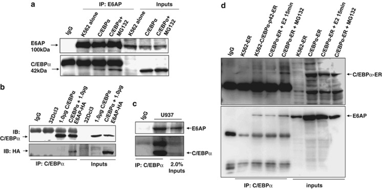
C/EBPα and E6AP physically interact in vivo in myeloid cells. (a) K562 cells were transiently transfected with C/EBPα and 24 h post transfection in one of the conditions cells were treated with 25 μM MG132 prior to cell lysate preparation. Co-immunoprecipitation using E6AP antibody was performed and the blot was probed with C/EBPα followed by E6AP antibody after stripping the same blot. (b) Co-immunoprecipitation using C/EBPα antibody was performed in lysates prepared from C/EBPα and E6AP co-transfected 32Dcl3 murine cells as indicated. Co-immunoprecipitates were resolved on 8% SDS-PAGE and probed with E6AP antibody followed by C/EBPα antibody after stripping the same blot. (c) Endogenous C/EBPα was co-immunoprecipitated from 2.5 mg whole cell lysates of U937 cells using C/EBPα antibody, resolved on 8% SDS-PAGE and probed with E6AP antibody followed by C/EBPα antibody after stripping the same blot. (d) K562-p42 C/EBPα-ER (C/EBPα-ER) stable cells were induced with 5 μM E2 for the indicated time points. Co-immunoprecipitation was performed with C/EBPα antibody. In one of the conditions 25 μM MG132 treatment was given for 3 h prior to cell lysate preparation. K562 cells transfected with empty vector were used as a control. The blot was probed with E6AP antibody. The same membrane was stripped and probed with C/EBPα antibody. Results are representative of minimum three independent experiments

