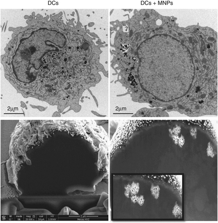
Figure 1.
TEM (upper images) and dual beam etched micrographs (lower images) of DCs without MNPs as the reference sample and DCs incubated with MNPs at 50 μg Fe3O4/ml. MNPs can be observed, as black particles in TEM images and as bright particles in dual beam images, within endocytic vesicles in the cytoplasmatic region

