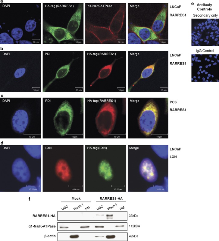
Figure 4.
RARRES1 is located in the ER, and LXN is located in the nucleus of prostate epithelial cell lines. Confocal immunofluorescence images depicting the location of HA-tagged RARRES1 in LNCaP (a, b) and PC3 (c) cells and LXN in LNCaP (d) cells 24 h after transfection. Cells we co-stained with anti-HA tag and (a) anti-α1-Na+/K+-ATPase (plasma membrane marker), (b, c) anti-protein disulphide isomerize (PDI; ER marker), (d) anti-LXN antibodies. Cells were counterstained with 4′-6-diamidino-2-phenylindole (DAPI) to enable nuclear visualization. White scale bar represents 10 μm. (e) Antibody controls using rabbit or mouse IgG instead of primary antibody and secondary antibody only. (f) Western blot data showing protein levels of RARRES1-HA (33 kDa) in unbroken cell fraction (UBC), wash fraction (Wash 1) and plasma membrane fraction (PM) of PC3 cells transfected with HA-tagged RARRES1 or reagent-only control (Mock) for 24 h before lysing the cells. Blots were probed with anti-HA (Santa Cruz) primary antibodies and horseradish peroxidase-linked secondary antibodies (Cell Signalling). Antibodies against α1-Na/K-ATPase (AbCam) and β-actin (Sigma) were used as plasma membrane and cytoplasmic internal controls, respectively.

