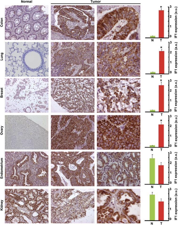
Figure 2.
IF1 is upregulated in some prevalent human carcinomas. Representative immunohistochemistries of IF1 expression in normal and tumor tissue of the colon, lung, breast, ovary, endometrium and kidney. Magnification × 20, × 40 and × 63. Histograms to the right of the pictures show the quantification of IF1 expression in normal (N, green, n=5) and tumor (T, red, n=10) specimens expressed as arbitrary units (a.u.). The results shown are the mean±s.e.m. *P<0.05 when compared with normal by Student's t-test. Note that whereas normal epithelial cells from the colon, lung, breast and ovary show low or negligible expression of IF1, endometrial and kidney cells have very-high expression of IF1.

