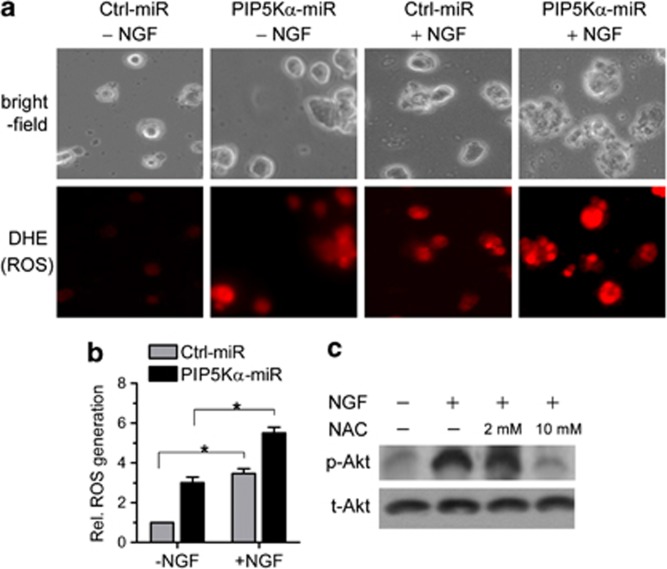
Figure 4.
Nerve growth factor (NGF)-induced reactive oxygen species (ROS) generation and its effect on Akt phosphorylation. (a) Control and PIP5Kα KD cells were incubated in the presence of 5 μℳ dihydroethidium (DHE), a fluorescent probe for ROS, for 20 min and then washed out with phosphate-buffered saline. Cells were further treated with or without NGF (100 ng ml−1) for additional 10 min. The red fluorescence of the probe owing to its oxidation was monitored using fluorescent microscopy. Cells were visualized on bright field channel. LED channel. (b) The fluorescent intensities of DHE were determined by image analysis and quantified as fold-change over those in unstimulated control cells. Values are mean±s.e.m. *P<0.01 (c) PIP5Kα KD cells were pretreated with N-acetyl-ℒ-cysteine (NAC) for 30 min and then treated with or without NGF (100 ng ml−1) for 15 min as indicated. Changes in phosphorylated and total levels of Akt were measured by western blotting.

