Figure 5.
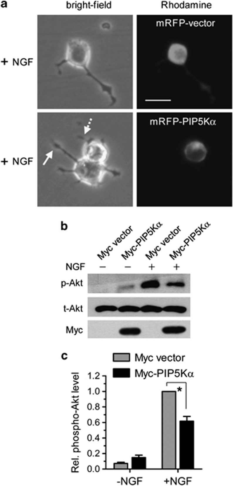
Effects of reconstituted PIP5Kα expression on nerve growth factor (NGF)-induced neurite outgrowth and Akt phosphorylation. PIP5Kα KD cells were transfected with monomeric red fluorescence protein (mRFP)–PIP5Kα (a) or Myc–PIP5Kα (b) for 24 h. mRFP or Myc empty vector was transfected as a corresponding control. (a) Following NGF treatment for 24 h, neurite outgrowth (bright field channel) and mRFP expression (Rhodamine channel) were detected by fluorescence microscopy. Note the difference in neurite length between the mRFP–PIP5Kα non-transfected cell (straight line arrow) and mRFP–PIP5Kα-transfected cell (dotted line arrow). Scale bar, 20 μm. (b) After treatment with or without NGF for 15 min, Akt phosphorylation was analyzed by western blotting. Myc–PIP5Kα expression was ascertained by anti-Myc western blot. (c) The phosphorylation levels of Akt in (b) were calculated as fold-change over that in NGF-stimulated vector-transfected condition. Values are mean±s.e.m. *P<0.01.

