Figure 6.
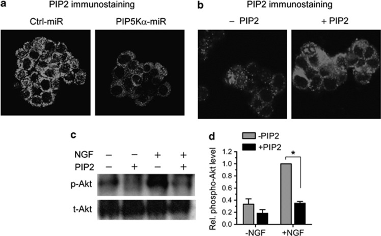
Effect of phosphatidylinositol (PI) 4,5-bisphosphate (PIP2) delivery on PIP2 levels and nerve growth factor (NGF)-induced Akt phosphorylation. (a) Control and PIP5Kα KD cells were examined for PIP2 levels by PIP2 immunostaining with a PIP2-specific antibody. Cells were further stained with biotin-labeled secondary antibody and then with Alexa Fluor 594-conjugated streptavidin. The resulting immune complexes were visualized by fluorescence microscopy. (b and c) PIP5Kα KD cells were preincubated with an equimolar complex of PIP2 and histone (final 10 μℳ each, +PIP2) or with histone only (−PIP2) for 1 h. (b) Changes in PIP2 levels were assayed by the PIP2 imaging in the same manner as described in (a). (c) Cells were further treated with or without NGF (100 ng ml−1) for 15 min under the indicated conditions. Akt phosphorylation was measured by western blot analysis. (d) The Akt phosphorylation levels in (c) were calculated as fold change over that in NGF-stimulated condition without PIP2. Values are mean±s.e.m. *P<0.01. miRNA, microRNA.

