Figure 1.

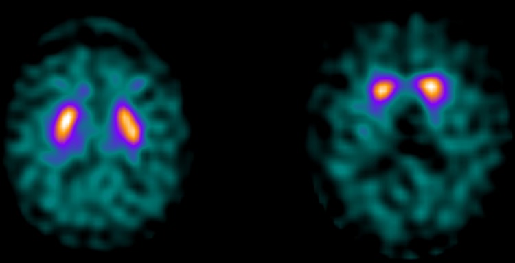
Transversal (123I)FP-CIT SPECT image from a patient in the dementia with Lewy bodies (DLB) cluster with a normal scan (left: case 2) and a patient with an abnormal scan in the non-DLB cluster (right: case 4).

Transversal (123I)FP-CIT SPECT image from a patient in the dementia with Lewy bodies (DLB) cluster with a normal scan (left: case 2) and a patient with an abnormal scan in the non-DLB cluster (right: case 4).