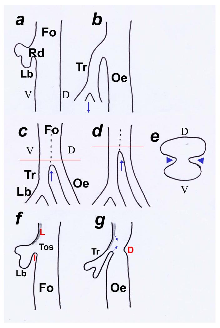
Fig. 1. Models of tracheo-oesophageal separation.
Schematic representation of the foregut illustrating theories of tracheo-oesophageal separation. Sagittal sections of the foregut (Fo) in a, b, c, d, f and g and transverse section in e at levels indicated in c and d. (a,b) One theory considers the respiratory diverticulum (Rd) to appear as a ventral evagination from the foregut with the two lung buds (Lb) at its caudal limit. It has been postulated that the trachea becomes separated from the oesophagus as a result of rapid downward growth (arrow in b) of the respiratory diverticulum. According to this theory, the trachea is never part of an undivided foregut and this model has been likened to a column of water emerging from a tap (tap and water theory). (c-e) An alternative theory suggests that the foregut initially elongates as an undivided structure having both ventral (V; tracheal) and dorsal (D; oesophageal) components (dotted line demarcates components in c and d). This theory suggests that the foregut then separates into the trachea (Tr) and oesophagus (Oe) as a result of the growth, in the coronal plane, of lateral mesenchymal ridges (arrowheads in e) which fuse to form a mesenchymal septum. Separation initially occurs at the level of the origin of the lung buds (Lb) and progresses in a rostral direction (arrows in c and d). A parallel theory supports the caudo-rostral progression of separation although it postulates that the lateral walls collapse and fuse, resulting in separation. This theory rejects the development of a septum. (f,g) In a third theory, paired laryngeal (L) and single inferior (I) folds define the tracheo-oesophageal space (Tos). Subsequent approximation (arrows) of these folds defines the separate trachea and oesophagus. The dorsal (D) fold marks the boundary between the pharynx and oesophagus.

