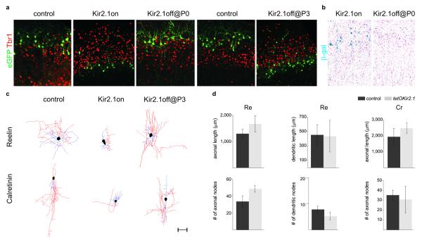
Figure 3. Specific interneuron subtypes require activity for migration and morphological maturation at two distinct stages of development.
a. Laminar positioning of P8 electroporated interneurons in wild type mice (control) and tetO-Kir2.1.ires.LacZ mice both co-electroporated with Dlx5/6-Tta and Dlx5/6-eGFP plasmids at e15.5. Mice received either no treatment (Kir2.1on); or were treated with Dox at e16.5 (Kir2.1off @ P0 onwards); or with Dox at P0 (Kir2.1off @ P3 onwards). b. β-galactosidase activity in P8 tetO-Kir2.1.ires.LacZ mice co-electroporated with Dlx5/6-Tta and Dlx5/6-eGFP plasmids either untreated or treated with Dox at e16.5 (Kir2.1off @ P0 onwards). c. Neurolucida reconstructions of Cr+ and Re+ interneurons in wild type (control) and tetO-Kir2.1.ires.LacZ mice both co-electroporated with Dlx5/6-Tta and Dlx5/6-eGFP plasmids. Mice received either no Dox treatment (Kir2.1on) or Dox at P0 (Kir2.1off @ P3 onwards). Axons are shown in red, dendrites in blue and somata in black. Scale bar: 50 μm d. Quantification of dendritic and axonal morphology in control and experimental Cr+ and Re+ interneurons in tetO-Kir2.1.ires.LacZ mice after Dox administration at P0. Mean percentage values (±SEM) were obtained from >3 reconstructed interneurons each in Dox-treated wild type and tetO-Kir2.1.ires.LacZ mice for each subtype analyzed at P8.

