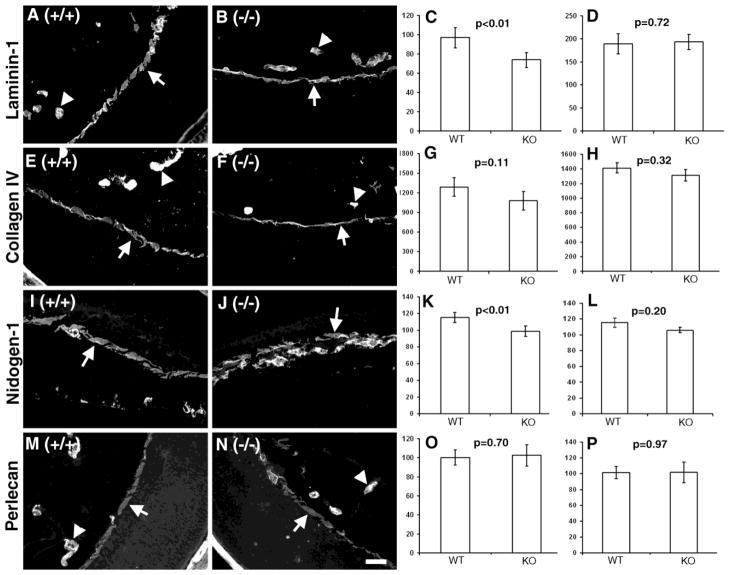
Figure 6. Laminin-111 and nidogen-1 were reduced in POMGnT1 knockout ILM.
Eye sections of wildtype (A, E, I, and M) and POMGnT1 knockout mice (B, F, J, and N) were stained with antibodies against laminin-111 (A and B), collagen IV (E and F), nidogen-1 (I and J), and perlecan (M and N). The fluorescent intensity per μm2 area was measured and quantified for ILM (C, G, K, and O) and blood vessels (D, H, L, and P). Note that laminin-111 and nidogen-1 were significantly reduced in POMgnT1 knockout ILM when compared to the wildtype. Arrows: ILM. Arrowheads: Blood vessels. Scale bar in N: 2 μm.

