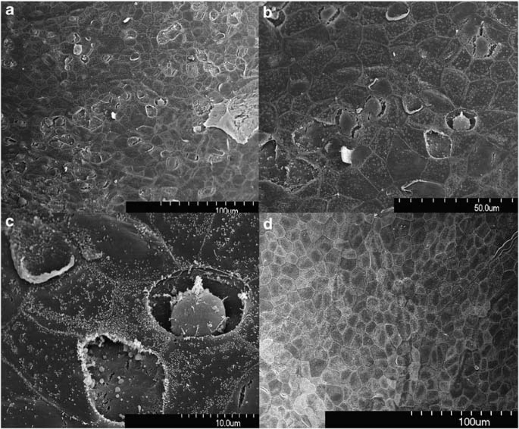
Figure 2.
SEM view of the epithelial surface facing the tympanic cavity. Samples taken 1 h after enzyme treatment (a–c from one sample with different magnification) show damaged and lost epithelia. Shrinkage of cells resulted in separation from surrounding cells. RWM from the control animal and at 3–4 weeks after the digestion show no signs of damage (d).

