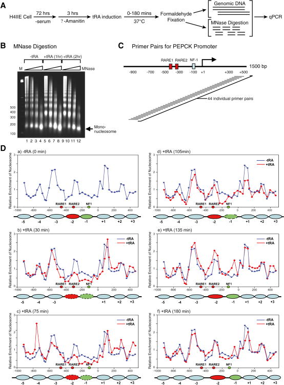
Figure 6.
Dynamic nucleosome repositioning at the PEPCK promoter during transcription activation in vivo.
(A) Protocol used for nucleosomal mapping in vivo. (B) Microccocal nuclease digestion of chromatin isolated from H4IIE cells as a function of treatment with tRA. (C) A schematic of the tiling primers covering the PEPCK promoter used for identifying in vivo nucleosomal positions are shown. (D) The dynamic change in nucleosomal positions at the PEPCK promoter in the absence (blue graph) and in the presence (red graph) of tRA induction (30-180 min). The filled ovals represent the nucleosomes covering RARE2 (red) and NF1 (green) that are repositioned upon tRA exposure.

