
Figure 1.
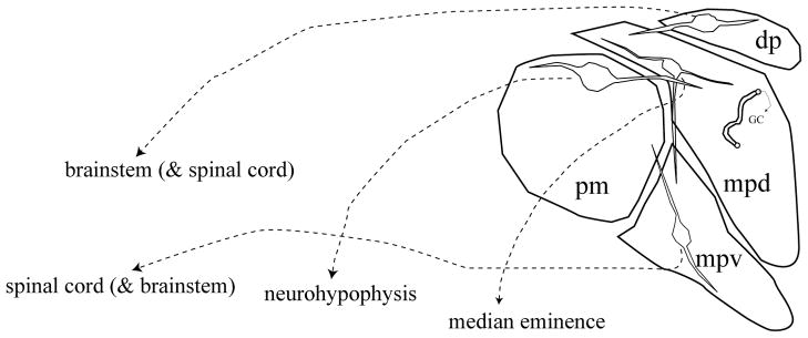
Schematic of the organization of the hypothalamic paraventricular nucleus. Neurons in the dorsal parvocellular (dp) and ventral division of the medial parvocellular regions (mpv) project primarily to brainstem and spinal cord regions associated with autonomic control. Neurons present in the dorsal division of the medial parvocellular zone (mpd) project primarily to the median eminence and control ACTH release. The posterior magnocellular (pm) subdivision sends projections to the neurohypophysis. Note that the dendrites of neurons localized in all three regions ramify across subdivisions, allowing for intranuclear communication.