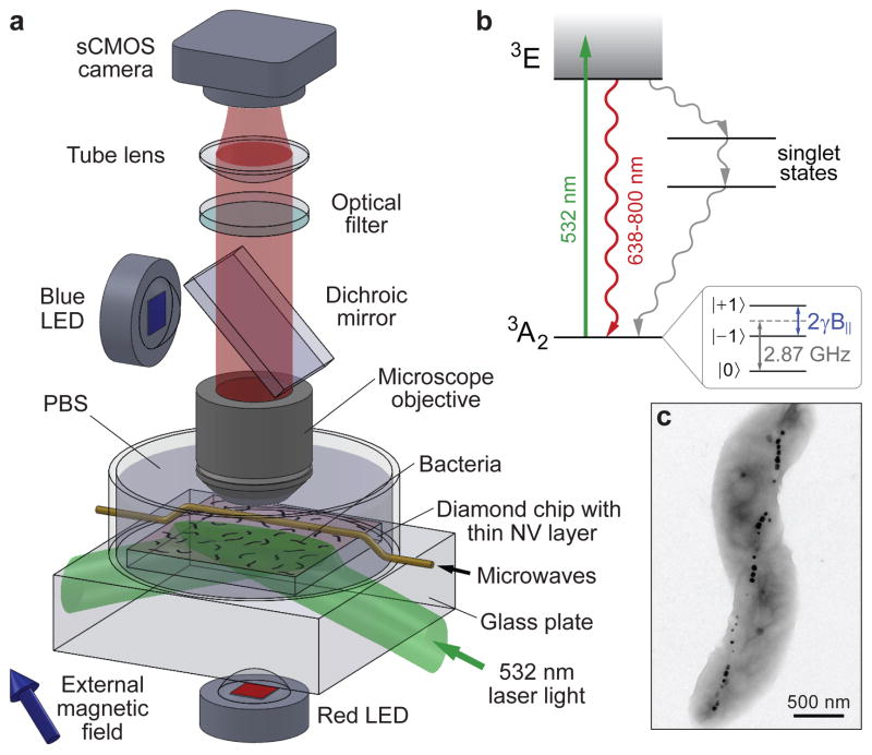
Figure 1. Wide-field magnetic imaging microscope.
a, Home-built wide-field fluorescence microscope used for combined optical and magnetic imaging. Live magnetotactic bacteria (MTB) are placed in phosphate-buffered saline (PBS) on the surface of a diamond chip implanted with nitrogen vacancy (NV) centres. Vector magnetic field images are derived from optically detected magnetic resonance (ODMR)8–10 interrogation of NV centres excited by a totally-internally-reflected 532 nm laser beam, and spatially correlated with bright field optical images. b, Energy-level diagram of NV centre; see Methods for details. c, Typical transmission electron microscope (TEM) image of a Magnetospirillum magneticum AMB-1 bacterium. Magnetite nanoparticles appear as spots of high electron density.

